
The human heart
How the heart is built, how blood flows through it, and how to draw and label it for your Class 10 exam, with Class 11 notes.
By Rahul Sain · Updated Nov 3, 2026
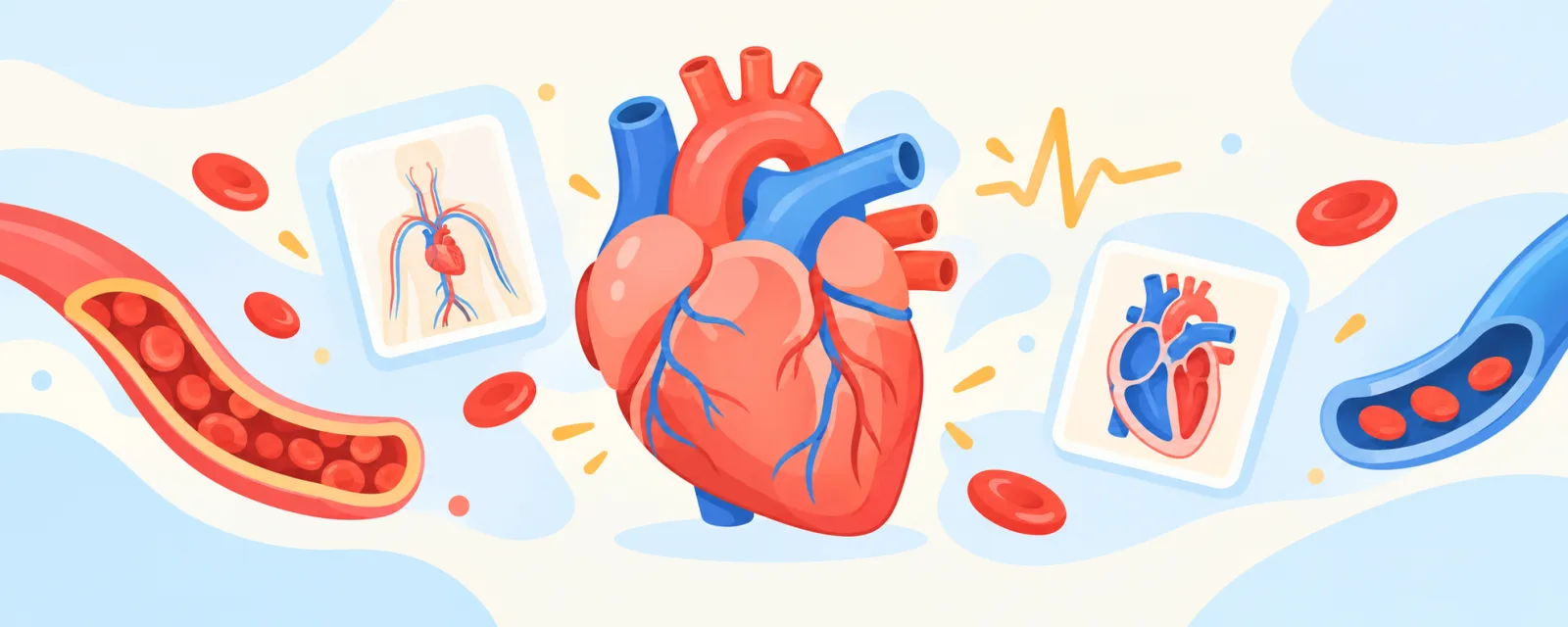 Free 3D labTry it in the 3D labHuman heart: take the readings yourself, free, no sign-in needed.
The human heart is a muscular pump about the size of your fist, with four chambers: two thin-walled atria on top that receive blood and two thick-walled ventricles below that pump it out. The right side sends deoxygenated blood to the lungs, and the left side sends oxygenated blood to the whole body, so blood passes through the heart twice in each full trip round the body. That is double circulation.
This guide covers what Class 10 asks: structure, every diagram label, the path of blood, the heartbeat, blood pressure and the "why" questions. Sections marked Class 11 add the cardiac cycle, cardiac output, the conducting system and the ECG.
Quick answers
- How many chambers are there in the human heart? Four: right atrium, right ventricle, left atrium, left ventricle.
- Valves: let blood flow one way only. Tricuspid (right atrium to right ventricle), bicuspid or mitral (left atrium to left ventricle), and the pulmonary and aortic semilunar valves at the two exits.
- Path of blood: body → venae cavae → right atrium → right ventricle → pulmonary artery → lungs → pulmonary veins → left atrium → left ventricle → aorta → body.
- Double circulation: blood goes through the heart twice per cycle: once for the lungs (pulmonary circulation), once for the body (systemic circulation).
- Thickest wall: the left ventricle, because it pumps blood to the whole body.
- Blood pressure: normal is about 120/80 mm of Hg: 120 systolic, 80 diastolic.
- The exceptions: the pulmonary artery carries deoxygenated blood; the pulmonary veins carry oxygenated blood.
Where the heart is in your syllabus
- CBSE Class 10 (2026-27): Unit II, World of Living: "Basic concept of nutrition, respiration, transport and excretion in plants and animals" (CBSE Science X 2026-27).
- NCERT Class 10: Chapter 5, Life Processes (Reprint 2026-27), section 5.4.1 "Transportation in Human Beings", with Figure 5.10, the "Schematic sectional view of the human heart" you must draw (NCERT Science X, Ch. 5).
- CBSE / NCERT Class 11: Chapter 15, Body Fluids and Circulation: structure of the heart, cardiac cycle, cardiac output, ECG, double circulation and regulation of cardiac activity (CBSE Biology 2026-27, NCERT Biology XI, Ch. 15).
- ICSE Class 10 (2027): "Structure and working of the heart" with the main blood vessels entering and leaving it, "Concept of systole and diastole; concept of double circulation", and "Brief idea of pulse and blood pressure". Practical work includes identifying the heart's internal structure through models and charts (CISCE ICSE Biology 2027).
Structure of the human heart
The heart sits in the chest cavity between the two lungs, slightly tilted to the left. It is wrapped in a double-walled bag, the pericardium, which holds pericardial fluid, and it is made entirely of cardiac muscle (NCERT Biology XI, Ch. 15). NCERT Class 10 explains why it has separate chambers: blood carries both oxygen and carbon dioxide, and the chambers stop oxygen-rich blood mixing with blood that contains carbon dioxide (NCERT Science X, Ch. 5).
- Atria: two smaller, thin-walled upper chambers that receive blood.
- Ventricles: two larger lower chambers with thick muscular walls that pump blood out.
- Septum: the dividing wall. Class 11 names the thin inter-atrial septum, the thick inter-ventricular septum, and the fibrous atrio-ventricular septum, whose openings are guarded by valves.
- Valves: tricuspid and bicuspid between the atria and ventricles, and two semilunar valves at the exits. They allow flow "only in one direction" (NCERT Biology XI, Ch. 15).
- Vessels: venae cavae and pulmonary veins bring blood in; the pulmonary artery and aorta take it out.
How to draw the labelled diagram of the heart (Class 10)
The diagram you need is NCERT Figure 5.10, "Schematic sectional view of the human heart". Questions are often worded "Draw a sectional view of the human heart and label the following".
- Outline: a rounded shape, wider at the top, narrowing to the apex at the bottom, pointing slightly to the right of your page.
- Septum: a thick wall down the middle to near the apex. The heart's right half is on the left of your page.
- Chambers: a line across each half about one-third of the way down. Atria above, ventricles below.
- Wall thickness: atria thin, right ventricle medium, left ventricle clearly thickest. Examiners notice this.
- Valves: flaps where each atrium opens into its ventricle, pointing down, and small cup-shaped flaps at the bases of the pulmonary artery and aorta.
- Vessels: venae cavae into the top and bottom of the right atrium; pulmonary artery out of the right ventricle, splitting in two; pulmonary veins into the left atrium; aorta out of the left ventricle, arching over the top.
- Arrows for blood flow; if colour is allowed, the right side blue and the left side red.
- Labels with straight horizontal lines that do not cross, and the title.
The rule that loses most marks: the diagram is drawn as if you are facing the person, so the heart's right side is on the left of your page. NCERT Figure 5.10 is drawn this way.
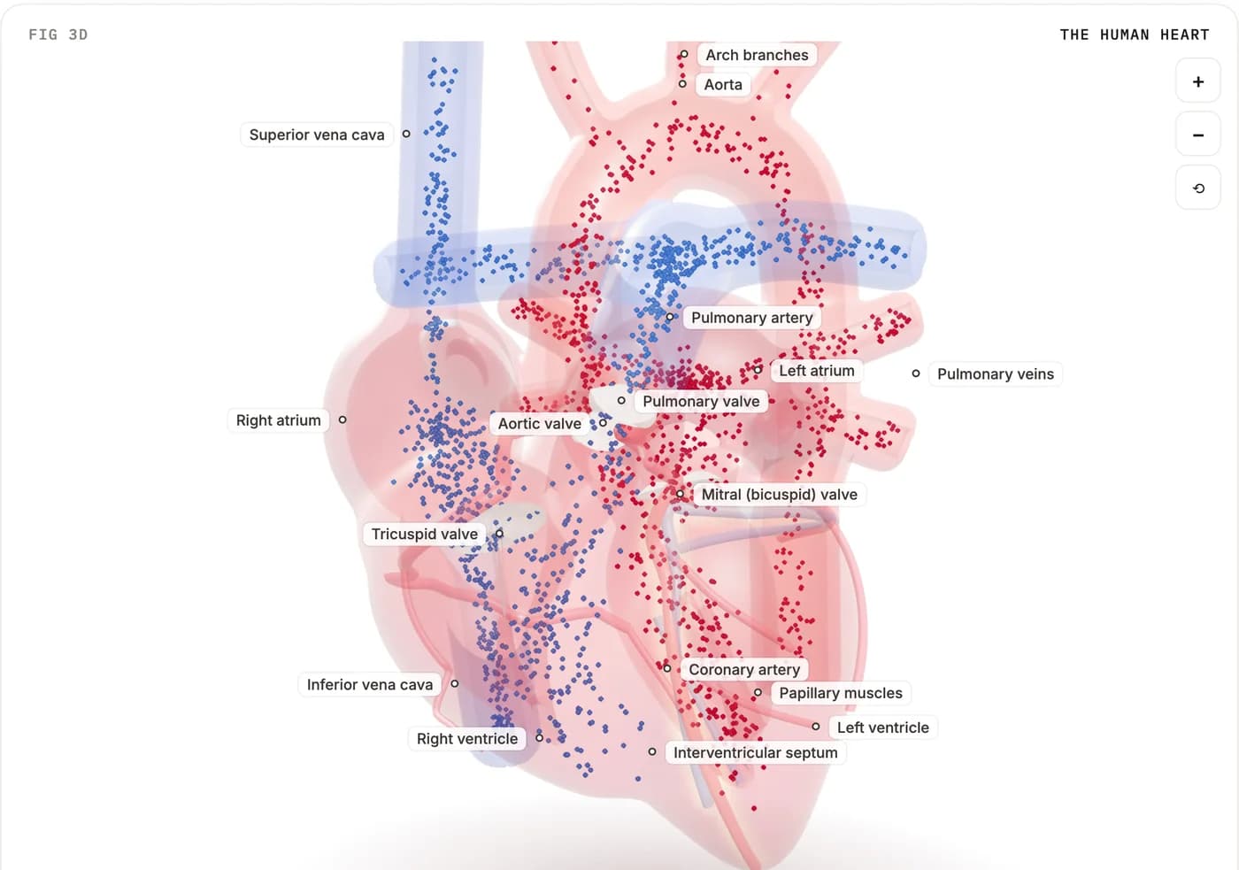
Every label you can be asked, and where it goes
"Class 10" means the label is on NCERT Figure 5.10. "Class 11" means it is in the text or Figure 15.2 of NCERT Biology XI, Chapter 15. "Beyond NCERT" labels appear in reference books and on our 3D heart, so know them, but they are not in the NCERT figures.
| Label | Where it is | What it does | Needed for |
|---|---|---|---|
| Right atrium | Upper right (left of your page) | Receives deoxygenated blood from the venae cavae | Class 10 |
| Right ventricle | Lower right | Pumps deoxygenated blood into the pulmonary artery | Class 10 |
| Left atrium | Upper left | Receives oxygenated blood from the pulmonary veins | Class 10 |
| Left ventricle | Lower left; thickest wall | Pumps oxygenated blood into the aorta | Class 10 |
| Septum (dividing wall) | Down the middle | Keeps the two kinds of blood apart | Class 10 |
| Interventricular septum | Between the ventricles | Thick muscular part of the septum | Class 11 |
| Tricuspid valve | Right atrium to right ventricle; three cusps | Stops backflow into the atrium | Class 10 as "valves"; named in Class 11 |
| Bicuspid (mitral) valve | Left atrium to left ventricle; two cusps | Stops backflow into the atrium | Class 10 as "valves"; named in Class 11 |
| Pulmonary valve (semilunar) | Exit of the right ventricle | Stops backflow into the ventricle | Class 11 |
| Aortic valve (semilunar) | Exit of the left ventricle | Stops backflow into the ventricle | Class 11 |
| Chordae tendineae | Cords from the tricuspid and mitral flaps to the ventricle wall | Hold the flaps so they cannot flip back | Class 11 (Fig. 15.2) |
| Papillary muscles | Inner ventricle wall, attached to the chordae | Tighten the chordae so the flaps are not forced into the atria | Beyond NCERT |
| Superior vena cava | Top of the right atrium | Brings blood from above the diaphragm: head, neck, arms, chest | Class 10 ("vena cava from upper body") |
| Inferior vena cava | Bottom of the right atrium | Brings blood from below the diaphragm: legs, abdomen | Class 10 ("vena cava from lower body") |
| Pulmonary artery | From the right ventricle; the trunk splits into right and left pulmonary arteries | Carries deoxygenated blood to the lungs | Class 10 ("pulmonary arteries") |
| Pulmonary veins | Four veins entering the left atrium | Bring oxygenated blood from the lungs | Class 10 |
| Aorta | From the left ventricle, curving over as the aortic arch | Carries oxygenated blood to the body | Class 10 |
| Arch branches | Brachiocephalic, left common carotid and left subclavian arteries | Supply the head, neck and arms | Beyond NCERT |
| Coronary arteries | From the first part of the aorta, over the heart's surface | Supply the heart muscle itself | Class 11 (text) |
| Sino-atrial node (SAN) | Right upper corner of the right atrium | Pacemaker: starts each beat | Class 11 |
| Atrio-ventricular node (AVN) | Lower left corner of the right atrium | Passes the signal on to the ventricles | Class 11 |
| Bundle of His (AV bundle) | From the AVN down the septum, splitting into right and left bundles | Carries the signal down to the ventricles | Class 11 |
| Purkinje fibres | Throughout both ventricle walls | Spread the signal so the ventricles contract | Class 11 |
| Apex | Bottom tip of the heart | — | Class 11 (Fig. 15.2) |
Sources: NCERT Science X, Fig. 5.10; NCERT Biology XI, Fig. 15.2 and 15.3.1; OpenStax 19.1 for cusps, chordae, papillary muscles, venae cavae, pulmonary trunk and veins, and coronary arteries; OpenStax 20.5 for the aortic arch. Our 3D heart uses the same names, with "Mitral valve" for the bicuspid valve.
Path of blood through the heart
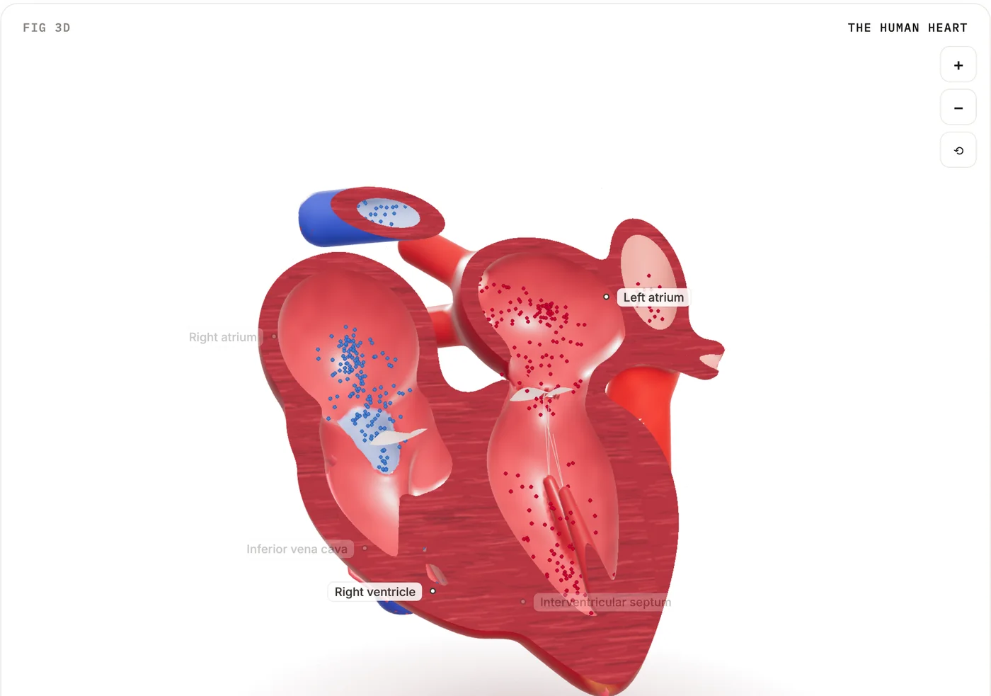
NCERT describes the flow step by step (NCERT Science X, Ch. 5). The two sides work at the same time.
Right side (deoxygenated blood, to the lungs):
- Deoxygenated blood from the body enters the right atrium through the superior and inferior venae cavae while the right atrium relaxes.
- The right atrium contracts and the right ventricle relaxes. Blood passes through the tricuspid valve into the right ventricle.
- The right ventricle contracts and pumps blood through the pulmonary valve into the pulmonary artery, to the lungs.
- In the lungs the blood takes in oxygen and gives up carbon dioxide.
Left side (oxygenated blood, to the body):
- Oxygenated blood from the lungs comes through the pulmonary veins into the left atrium while it relaxes.
- The left atrium contracts and the left ventricle relaxes. Blood passes through the bicuspid (mitral) valve into the left ventricle.
- The muscular left ventricle contracts and pumps blood through the aortic valve into the aorta.
- The aorta's branches carry the blood to every organ. Deoxygenated blood returns through veins to the venae cavae, and the cycle repeats.
The NHLBI gives the same route and valves (NHLBI).
Double circulation in human beings
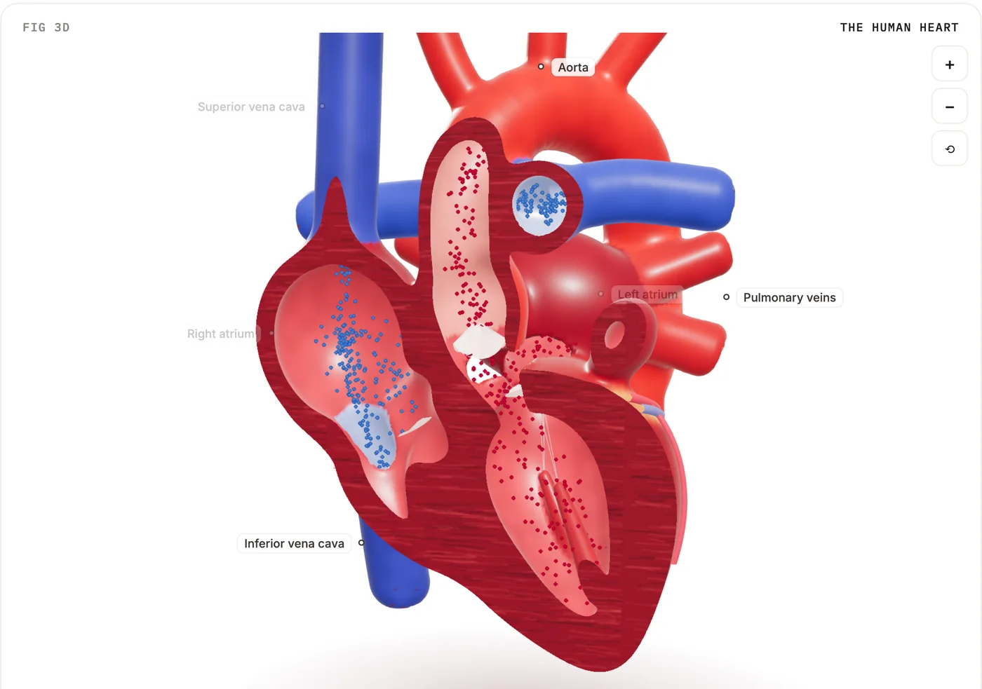
What it is. In one complete circuit, blood passes through the heart twice. NCERT: blood goes through the heart only once in a fish during one cycle, but twice in other vertebrates, and this is double circulation (NCERT Science X, Ch. 5). Class 11 names the two loops (NCERT Biology XI, Ch. 15):
- Pulmonary circulation: right ventricle → pulmonary artery → lungs → pulmonary veins → left atrium.
- Systemic circulation: left ventricle → aorta → arteries, arterioles, capillaries → venules, veins, venae cavae → right atrium.
Why double circulation is necessary in human beings. Separating the right and left sides stops oxygenated and deoxygenated blood mixing, which gives a highly efficient supply of oxygen. Birds and mammals need this because they have high energy needs: they constantly use energy to keep their body temperature steady (NCERT Science X, Ch. 5).
Compared with other animals (NCERT Biology XI, Ch. 15):
| Animal | Heart | Circulation |
|---|---|---|
| Fish | 2 chambers (one atrium, one ventricle) | Single: heart → gills → body → heart |
| Amphibians, most reptiles | 3 chambers (two atria, one ventricle) | Incomplete double: blood mixes in the one ventricle |
| Crocodiles, birds, mammals | 4 chambers | Double: no mixing |
Working of the human heart: the heartbeat in simple terms
Systole means contraction; diastole means relaxation. One heartbeat is one cardiac cycle:
- The atria contract and push the last of their blood into the relaxed ventricles.
- The ventricles contract. The tricuspid and bicuspid valves shut so blood cannot go back into the atria: the first sound, "lub". The semilunar valves open and blood goes out into the pulmonary artery and aorta.
- The ventricles relax. The semilunar valves shut so blood cannot fall back into the ventricles: the second sound, "dub".
- All chambers relax and fill from the veins, and the next beat begins.
Class 10 NCERT says only that valves stop blood flowing backwards; the lub-dub detail is Class 11 (NCERT Biology XI, Ch. 15).
Pulse and blood pressure
Blood pressure is the force that blood exerts against the wall of a vessel. It is much greater in arteries than in veins. The pressure in an artery during ventricular systole is the systolic pressure, and during ventricular diastole the diastolic pressure: normally about 120 and 80 mm of Hg, written 120/80. It is measured with a sphygmomanometer. High blood pressure is called hypertension; NCERT says it is caused by the constriction of arterioles, which increases resistance to blood flow (NCERT Science X, Ch. 5).
Pulse (ICSE asks for a brief idea). Each time the left ventricle pushes blood into the aorta, the elastic artery walls stretch and then recoil. This expansion and recoil is the pulse (OpenStax A&P 2e, 20.2). You feel it where an artery is close to the skin, usually the wrist below the thumb, or the neck. Beats per minute give the pulse rate, which equals the heart rate. For adults and children aged 10 and older, a normal resting rate is 60 to 100 beats per minute (MedlinePlus).
Class 11: the cardiac cycle, phase by phase
NCERT Biology XI describes the cycle as a fixed sequence of systole and diastole of the atria and ventricles (NCERT Biology XI, Ch. 15):
- Joint diastole. All four chambers are relaxed. The tricuspid and bicuspid valves are open, so blood from the pulmonary veins and venae cavae flows through the atria into the ventricles. The semilunar valves are closed.
- Atrial systole. The SAN fires and both atria contract together. This increases the flow of blood into the ventricles by about 30 per cent.
- Ventricular systole. The signal reaches the ventricles through the AVN and the bundle of His, and they contract while the atria relax. Rising ventricular pressure shuts the tricuspid and bicuspid valves (lub). As it rises further, it forces the semilunar valves open and blood flows into the pulmonary artery and aorta.
- Ventricular diastole. The ventricles relax, their pressure falls, and the semilunar valves close (dub). As the pressure falls further, the tricuspid and bicuspid valves are pushed open by blood in the atria, and the ventricles fill again.
Physiology books split ventricular systole into isovolumetric contraction (all valves shut, pressure rising) and ventricular ejection, and ventricular diastole into isovolumetric relaxation and filling (OpenStax A&P 2e, 19.3). Our 3D heart shows these phase names live, with filling split into "Rapid filling" and "Diastasis (slow filling)".
Timings at 72 beats per minute
NCERT: the heart beats 72 times a minute, "the duration of a cardiac cycle is 0.8 seconds". Strictly 60 ÷ 72 = 0.83 s, which NCERT rounds to 0.8 s. NCERT does not split the cycle further. OpenStax 19.3 gives atrial systole as about 0.1 s and ventricular systole as about 0.27 s, which round to these figures for a 0.8 s cycle:
| Event | Time |
|---|---|
| Atrial systole | 0.1 s |
| Ventricular systole | 0.3 s |
| Joint diastole (all chambers relaxed) | 0.4 s |
| Whole cycle | 0.1 + 0.3 + 0.4 = 0.8 s |
From these: atrial diastole = 0.8 − 0.1 = 0.7 s, and ventricular diastole = 0.8 − 0.3 = 0.5 s. So both the atria and the ventricles spend longer relaxing than contracting.
Class 11: stroke volume and cardiac output
- Stroke volume: the blood each ventricle pumps out per beat, about 70 mL.
- Cardiac output: the volume each ventricle pumps per minute = stroke volume × heart rate. NCERT gives an average of 5000 mL, or 5 litres, a minute (NCERT Biology XI, Ch. 15).
Worked example. Stroke volume 70 mL, heart rate 72 beats/min:
Cardiac output = 70 mL × 72 /min = 5,040 mL/min = 5.04 L/min ≈ 5 L/min
Units check: mL per beat × beats per minute = mL per minute. Correct.
NCERT adds that the body can change both stroke volume and heart rate, so an athlete's cardiac output can be much higher than an ordinary person's.
Ejection (beyond NCERT, shown on our 3D heart). A resting adult's ventricle holds about 130 mL when full (OpenStax A&P 2e, 19.3). If it pumps out 70 mL, it keeps 130 − 70 = 60 mL, and the fraction ejected is 70 ÷ 130 = 0.54, or about 54%.
Class 11: the conducting system and a simple ECG
The beat starts inside the heart itself, in nodal tissue, a special kind of cardiac muscle that can generate signals without any outside stimulus (NCERT Biology XI, Ch. 15):
- The SAN, in the right upper corner of the right atrium, generates the most signals, 70–75 a minute, so it sets the rhythm. It is called the pacemaker.
- The signal spreads over both atria, which contract.
- The AVN, in the lower left corner of the right atrium, picks it up and passes it to the AV bundle (bundle of His). The signal takes about 0.1 s to cross the AV node (OpenStax A&P 2e, 19.2).
- The bundle runs through the septum and splits into right and left bundles, which end in Purkinje fibres spread through the ventricle walls.
- The ventricles contract from the signal carried by these fibres.
Because the heart muscle starts its own beat, the heart is called myogenic. A centre in the medulla oblongata can adjust it through the autonomic nervous system: sympathetic nerves raise the heart rate and the strength of contraction, parasympathetic nerves lower the rate, and adrenal medullary hormones can also raise cardiac output.
ECG (electrocardiogram): a graph of the heart's electrical activity during a cycle. A standard ECG uses three leads, one on each wrist and one on the left ankle. NCERT names three parts:
- P wave: depolarisation (electrical excitation) of the atria, which makes both atria contract.
- QRS complex: depolarisation of the ventricles, which starts ventricular contraction. Contraction begins shortly after Q and marks the start of systole.
- T wave: the ventricles returning to normal (repolarisation). The end of the T wave marks the end of systole.
Counting QRS complexes over a set time gives the heart rate. Worked example: 18 QRS complexes in 15 seconds → 18 × (60 ÷ 15) = 18 × 4 = 72 beats per minute.
Why the left ventricle has the thickest wall, and other "why" questions
Why is the left ventricle wall thicker than the right? The left ventricle pumps blood through the aorta to the whole body, a long circuit with high resistance, so it must produce a much higher pressure. The right ventricle pumps only to the nearby lungs, and the pulmonary circuit is shorter with less resistance (OpenStax A&P 2e, 19.1; NHLBI).
Why do ventricles have thicker walls than atria? NCERT: "Since ventricles have to pump blood into various organs, they have thicker muscular walls than the atria do." The atria only push blood into the ventricles just below them (NCERT Science X, Ch. 5).
Why do arteries have thick, elastic walls and veins have valves? Blood leaves the heart under high pressure. In veins it is no longer under pressure, so their walls are thinner, and valves keep it moving one way (NCERT Science X, Ch. 5).
Common mistakes in exams
- Swapping left and right in the diagram. Check: the venae cavae enter the chamber on your left, and the aorta leaves the thick chamber on your right.
- "Arteries carry oxygenated blood, veins carry deoxygenated blood." That is not the definition. Arteries carry blood away from the heart; veins bring it back.
- Forgetting the pulmonary exceptions. The pulmonary artery carries deoxygenated blood (right ventricle to lungs); the pulmonary veins carry oxygenated blood (lungs to left atrium).
- Swapping the valves. Tricuspid is on the right, bicuspid (mitral) on the left. Memory aid: "tRIcuspid on the RIght".
- Mixing up the heart sounds. "Lub" is the tricuspid and bicuspid valves closing; "dub" is the semilunar valves closing.
Human heart questions and answers (Class 10)
- Which chamber of the heart receives oxygenated blood from the lungs? The left atrium, through the pulmonary veins.
- Which chamber pumps oxygenated blood to the body? The left ventricle, through the aorta.
- Which blood vessel carries deoxygenated blood from the heart to the lungs? The pulmonary artery, from the right ventricle.
- Why is it necessary to separate oxygenated and deoxygenated blood in mammals and birds? (NCERT in-text question.) It gives a highly efficient oxygen supply, which they need because they use a lot of energy to keep their body temperature constant.
- Describe double circulation of blood in human beings. Why is it necessary? (NCERT exercise 11.) Blood passes through the heart twice per cycle: right ventricle → lungs → left atrium (pulmonary), then left ventricle → body → right atrium (systemic). It keeps the two kinds of blood apart, giving an efficient oxygen supply for our high energy needs.
- How does circulation in a fish differ from that in humans? A fish has a two-chambered heart and single circulation: heart → gills → body → heart, through the heart once per cycle.
- Define systolic and diastolic pressure and give their normal values. Systolic: arterial pressure during ventricular contraction, about 120 mm of Hg. Diastolic: during ventricular relaxation, about 80 mm of Hg.
- (Class 11) Calculate the cardiac output if the stroke volume is 70 mL and the heart rate is 72 per minute. 70 × 72 = 5,040 mL/min ≈ 5 L/min.
Try it in 3D
Student Intelligence has a free 3D human heart at /labs/human-heart. Turn and zoom it, and switch between four views: Whole, Four chambers, Front cut and X-ray (the cut views have a cut-depth slider).
Blood moves through it in blue (oxygen-poor) and red (oxygen-rich). Labels name the chambers, the four valves, the septum, the papillary muscles and the main vessels, and tapping a part (a chamber, valve, the septum, the papillary muscles or a coronary branch) tells you what it does. A heart-rate slider runs from 40 to 180 beats per minute; you can slow the beat to a tenth of normal speed, pause it or step through it.
Readouts show the phase, the left ventricle's volume and pressure, blood pressure, cardiac output, ejection, whether each valve is open or shut, and a strip of pressure, volume and ECG over one beat. Turn on Sound to hear a soft thump when the mitral valve shuts ("lub") and when the aortic valve shuts ("dub"). The numbers come from a computer model of a healthy adult at rest; the SAN and AVN are not drawn.
The 3D heart, its controls, and a short quiz and flashcards are free without an account. Asking the voice tutor questions needs a free sign-in.
FAQ
What is double circulation in human beings, and why is it necessary?
Blood passes through the heart twice in one complete circuit: once to the lungs and back, once to the body and back. It keeps oxygenated and deoxygenated blood apart, giving the efficient oxygen supply that birds and mammals need.
Which artery carries deoxygenated blood?
The pulmonary artery. It takes deoxygenated blood from the right ventricle to the lungs.
What causes the "lub-dub" sound of the heart?
Valves closing. "Lub" is the tricuspid and bicuspid valves shutting as the ventricles start to contract; "dub" is the semilunar valves shutting as the ventricles relax.
Is the heart diagram in Class 10 the same as in Class 11?
The chambers and vessels are the same. Class 10 uses NCERT Figure 5.10, a simple sectional view. Class 11 Figure 15.2 adds the SAN, AVN, bundle of His, chordae tendineae, interventricular septum and apex.
Want to see the four chambers and the valves working before you draw them? Open the free 3D human heart on Student Intelligence.
Sources
- NCERT. Science, Textbook for Class X, Chapter 5 "Life Processes" (Reprint 2026-27): section 5.4.1 Transportation in Human Beings, Figures 5.10 and 5.11, Blood pressure, blood vessels, in-text question 2 and exercise 11. https://ncert.nic.in/textbook/pdf/jesc105.pdf
- NCERT. Biology, Textbook for Class XI, Chapter 15 "Body Fluids and Circulation" (Reprint 2026-27): 15.3 Circulatory pathways, 15.3.1 Human circulatory system (Figure 15.2), 15.3.2 Cardiac cycle, 15.3.3 ECG, 15.4 Double circulation, 15.5 Regulation of cardiac activity. https://ncert.nic.in/textbook/pdf/kebo115.pdf
- CBSE. Curriculum 2026-27, Science, Class X: Unit II World of Living. https://cbseacademic.nic.in/web_material/CurriculumMain27/SecPart1/Science_SecP1_2026-27.pdf
- CBSE. Curriculum 2026-27, Biology, Classes XI–XII: Class XI Chapter 15 Body Fluids and Circulation. https://cbseacademic.nic.in/web_material/CurriculumMain27/SecPart2/Biology_SecP2_2026-27.pdf
- CISCE. ICSE 2027 Science (52) Biology syllabus: Class X, Human Anatomy and Physiology, Circulatory System; practical work. https://cisce.org/wp-content/uploads/2025/03/12.-Biology.pdf
- National Heart, Lung, and Blood Institute (NHLBI, NIH). How Blood Flows through the Heart. https://www.nhlbi.nih.gov/health/heart/blood-flow
- OpenStax. Anatomy and Physiology 2e, 19.1 Heart Anatomy. https://openstax.org/books/anatomy-and-physiology-2e/pages/19-1-heart-anatomy
- OpenStax. Anatomy and Physiology 2e, 19.2 Cardiac Muscle and Electrical Activity. https://openstax.org/books/anatomy-and-physiology-2e/pages/19-2-cardiac-muscle-and-electrical-activity
- OpenStax. Anatomy and Physiology 2e, 19.3 Cardiac Cycle. https://openstax.org/books/anatomy-and-physiology-2e/pages/19-3-cardiac-cycle
- OpenStax. Anatomy and Physiology 2e, 20.2 Blood Flow, Blood Pressure, and Resistance. https://openstax.org/books/anatomy-and-physiology-2e/pages/20-2-blood-flow-blood-pressure-and-resistance
- OpenStax. Anatomy and Physiology 2e, 20.5 Circulatory Pathways. https://openstax.org/books/anatomy-and-physiology-2e/pages/20-5-circulatory-pathways
- MedlinePlus (US National Library of Medicine). Pulse. https://medlineplus.gov/ency/article/003399.htm